Tesla’s Single Wire Shadowgraph Recreated
As with other high vacuum tubes recreated under Tesla’s name, the next step was to observe the production of speciality rays which Tesla dubbed as Shadowgraphs, since the fogging and exposures created on photographic plates resembled shadows. These results were observed by Tesla as early as 1894, but were not publicly announced until after Wilhelm C. Roentgen discovered X-Rays in 1896. It must be borne in mind to the reader that based upon Tesla’s observations as well as the author, the rays emanating from from such single terminal tubes are not solely X-Rays. For this purpose, the latter will be regarded as Roentgen Rays, and that of Tesla’s origin, Tesla Rays, as proper. This distinguishing factor will be examined in greater detail later in this article.
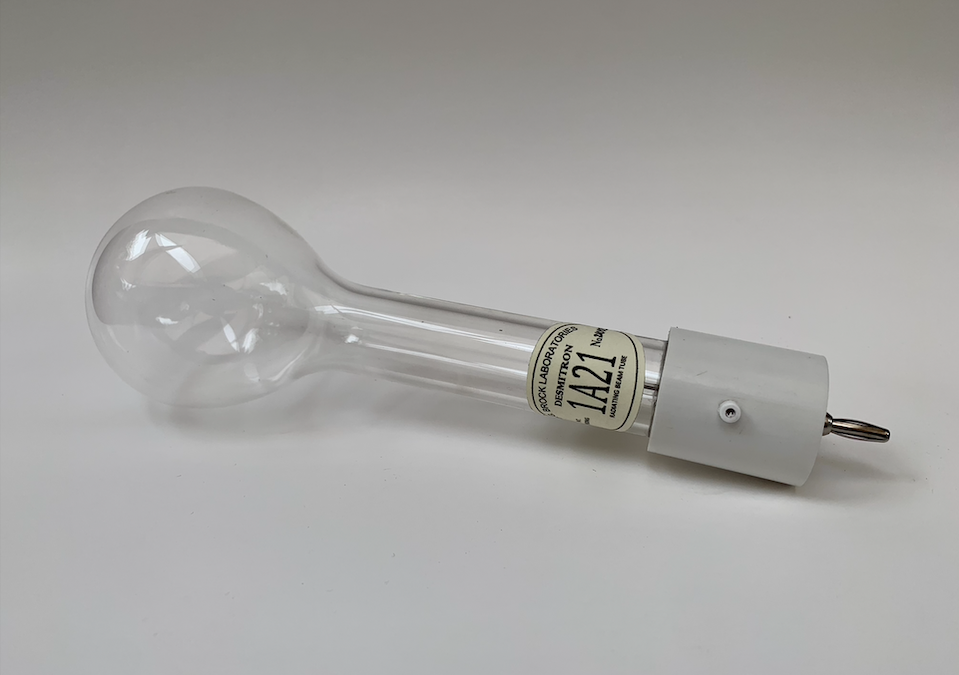
For the purpose of this initial experiment, Tesla’s original slender shadowgraph tube was not employed, whereby the tube’s envelope consists of a pear shaped glass. This decision was dependent upon the length of glass tubing available at the time, as the elongated version had required a greater length.
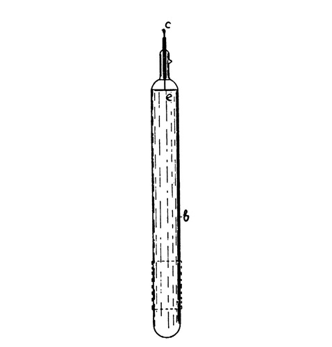
Fig. 1. – Tesla’s Shadowgraph Tube 
Fig. 2. – An experimental Shadowgraph Tube
Since previous shadowgraph tube experiments had failed to produce such rays in part to their ultra high vacuum being 10⁻⁸ Torr, the tube shown in figure (2), was evacuated to a degree of only 10⁻⁴ Torr. This proved to liberate a larger quantity of ions and project very strong rays in the direction in which the tube was pointed.
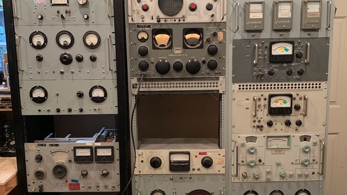
Rather than having the tube supplied with high potential direct current as is used with a typical Roentgen Tube, high frequency – high potential Alternate Currents were provided with a diathermy apparatus and series fed extra coil. This inductor served as to exemplify the rather feeble potential emanating from the apparatus, and magnify them by more than 100 fold.
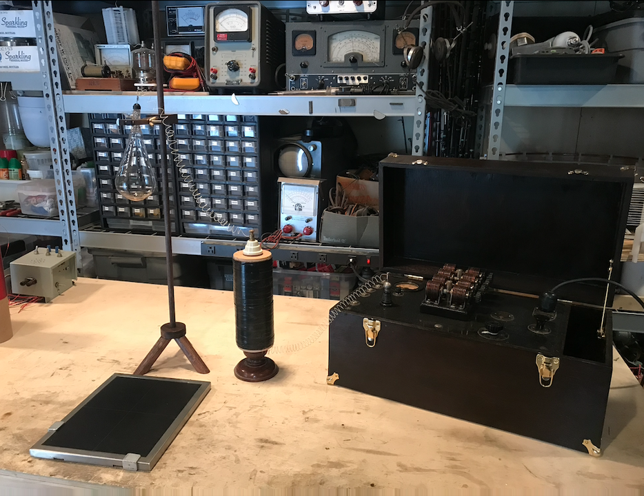
The above figure displays the operating condition for the tube, in which a photographic cassette is fixed in position so that to be inline with the emanating rays from the tube. Below is a closer view of the adjustable stand for the tube which allows it to project downwards, figure (4), or horizontally outwards, figure (5).
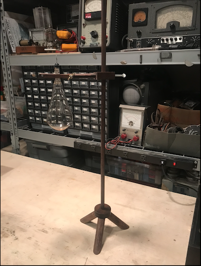
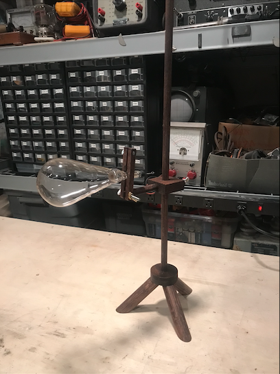
During operation, the tube produces magnificent sky blue and green hues around the glass, while a barely visible beam of matter appears to be issuing from the aluminum electrode. This beam is then projected onto the inner wall at the apex of the lamp, whereby an irregular blue patch of phosphorescence is observed. This is likely due to the projected matter physically bombarding a finite section of the glass, in turn producing a respectable quantity of heat in said region alone. Achievable photographs of the operation are shown in the following figures.
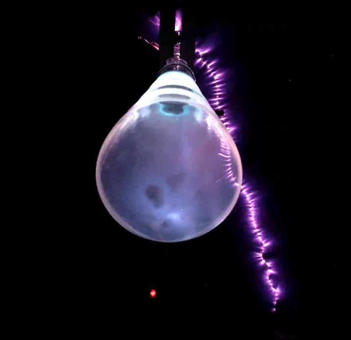
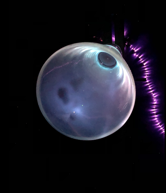
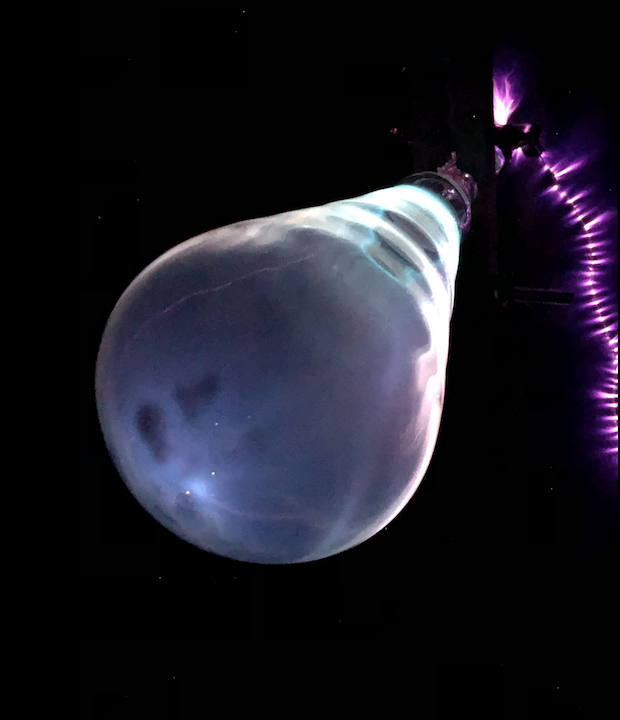
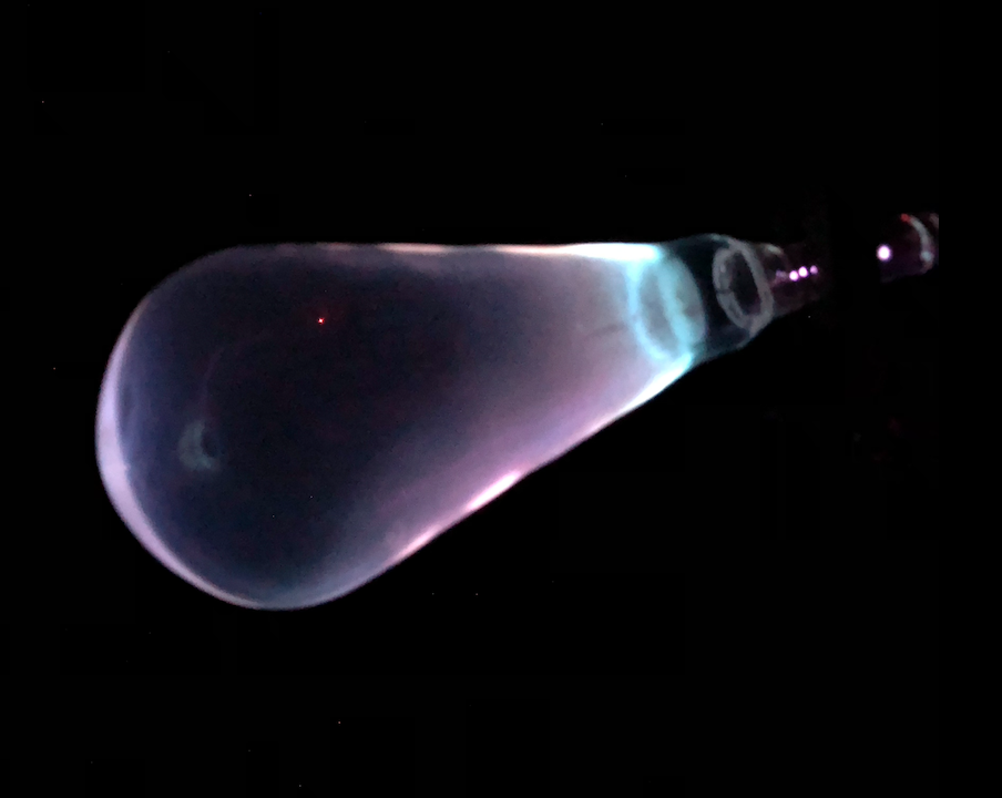
It must be noticed in figures (6) through (9) that the irregular patches of phosphorescence coincide with the projection of energized matter emanating off from the aluminum electrode, and concentrating at the apex of the lamp, longitudinal to the plane of the electrode. The intensity of the projected matter may be evident by inspection of the electrode, for which it possesses a defined spot of degradation, quintessential of the electrical removal of the of electrode’s material.

The resultant rays produced by this lamp were of immense nature, whereby a fluoroscope X-Ray intensifying screen allowed the interior of objects placed in front of the screen to be observed at a distance of seven feet away from the tube. Moreover, the rays emanating from the tube were clearly detected with a Geiger-Müller tube at a distance of 70 feet away, possibly extending themselves even further.
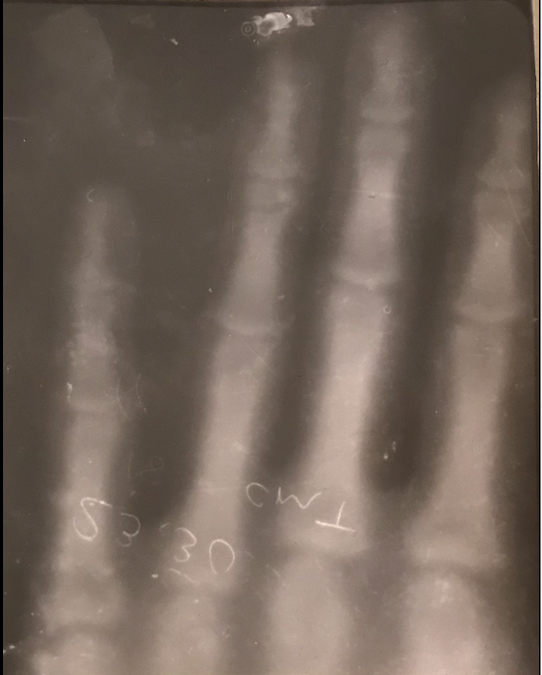
To that effect, the intensity of the rays allowed for the production of the following shadowgraph taken at a distance of six inches away from the tube. Note, that the clarity of the image suffers, as the determined focal length of this tube produces clear images at a minimum distance of 12 inches.
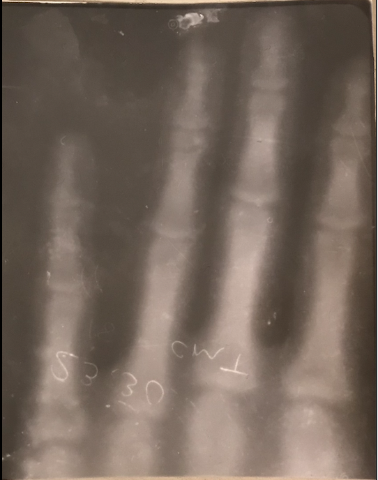
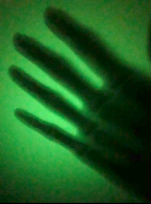

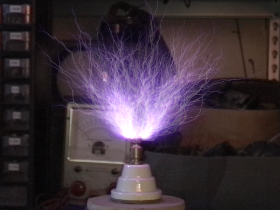
To the reader and concerned skeptic who wishes to denote these rays to be the working of X-Rays, it becomes worthy to note the minute differences associated with this rather unknown form of penetrating ray, the Tesla Ray. It appears that the electrostatic nature of these rays, rather than being electromagnetic as would be a conventional Roentgen Ray, allows for greater detection distances to be achieved. Along with the safety factor associated with such rays. Tesla remarks on the ability for these rays to be detected by photographic plates at a distance of 100 feet, suggesting a close proximity exposure, (if to be Roentgen Radiation), in excess of 50 Roentgens per hour. This exceedingly high level of exposure would entail detrimental and fatal health consequences over prolonged use. Tesla and his assistants had in fact endured many hours and days of experimentation with such rays, hence, some form of fatal cancer should have issued. To the dismay of history, this did not come to avail, as Mr. Tesla lived well into his eighties, and neither of his assistants during this period suffered adverse effects.
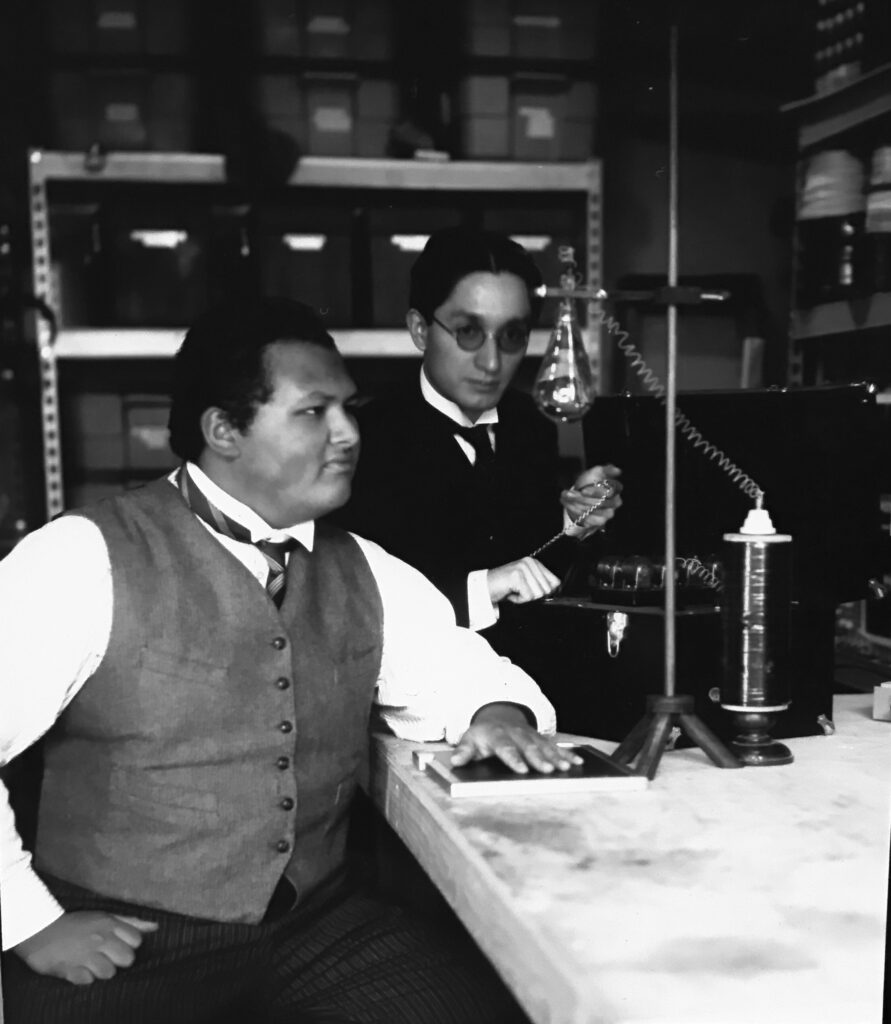
On the other hand, Thomas A. Edison, who was simultaneously exploring the world of Roentgen Rays with his assistant Clarence Madison Dally, had employed the typical two terminal cold cathode X-Ray tube in conjunction with a high potential Direct Current Apparatus to excite Roentgen Ray production. Despite the feeble power involved, Edison soon developed cataracts, meanwhile his assistant C.M. Dally perished, both being directly linked to their Roentgen Ray research.
Hence, it can be deduced that while Nikola Tesla manufactured enormous Alternate Currents to drive his Shadowgraph tubes and experienced no adverse health effects, Thomas Edison reported grave dangers associated with Roentgen Rays, produced by his two terminal slightly-high voltage fed tubes. A definite distinction may be arrived at by comparing these two avenues of research, for one appears to be a superior alternative.
In conjunction with Tesla’s reports, the author has deduced that in correlation with measurements of the detected rays at a distance of 70 feet to that of the Inverse Square Law, a close proximity exposure of one foot from the tube shown in figure (2) would entail a 4.5 Roentgen per hour exposure. This would ultimately exhibit noticeable physical damage to any nearby person within a few hours of working time. The reader will be surprised by the fact that there exists a lack of any abnormalities to the author, as well as individuals who partook in the experiments. Surely if the tube were producing high intensity Roentgen Rays, then negative effects would have occurred, when in fact this does not hold to be true in this circumstance. It is thus rationed that the Tesla Shadowgraph tube is in fact exhibiting unique features of a ray which does not conform to the classical Roentgen Ray. This is a new avenue of research which will be explored and expounded upon in upcoming relating articles.
Share this:
- Click to share on Twitter (Opens in new window)
- Click to share on Facebook (Opens in new window)
- Click to share on Pinterest (Opens in new window)
- Click to share on LinkedIn (Opens in new window)
- More
- Click to share on Reddit (Opens in new window)
- Click to share on WhatsApp (Opens in new window)
- Click to share on Mastodon (Opens in new window)
- Click to share on Telegram (Opens in new window)
- Click to share on Tumblr (Opens in new window)
- Click to share on Nextdoor (Opens in new window)
- Click to share on Pocket (Opens in new window)
- Click to email a link to a friend (Opens in new window)
